Newsletter

Tracheal Mites (Acarine)

Acarine eggs in tracheal tube. Courtesy The Animal and Plant Health Agency (APHA), Crown Copyrigh

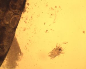
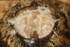
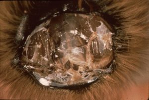
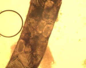
It is thought that the Tracheal mites (also known as Acarine) was a major contributory factor to the ‘Isle of Wight disease’ first seen in the early 1900s. It decimated the honey bee population, later spreading to mainland UK. In more recent times, the Tracheal mites have had a serious economic impact on the beekeeping industry in North America, after it’s introduction there in the 1980s from Mexico. However, in the UK Tracheal mite infection is not usually a serious disease, with relatively small numbers of colonies being affected.


The honey bee delivers oxygen to body tissues via diffusion through a complex system comprising of tubes called trachea and air sacs. It is in these trachea that the acarine mites reproduce and feed. Mature female mites enter the anterior thoracic spiracles of young bees (bees are only susceptible to infestation within the first nine days after emergence). The mites lay their eggs in the trachea (above) and upon hatching the larvae begin to feed on the haemolymph (blood) of the bee. The larvae undergo several moults before reaching their adult forms, and are then ready to infest new hosts.

Symptoms and Cause

Acarapisosis is the infestation of the breathing tubes (trachea) of the adult bee by the parasitic mite Acarapis woodi. In many cases bees cluster in front of the hive, appearing confused and

The tracheal mite, Acarapis woodi. Courtesy The Animal and Plant Health Agency (APHA), Crown Copyright

disorientated, unable to return to the hive. Some of the bees may also display what is known as ‘K-wings’, where the rows of hooks holding the pairs of the bee’s wings together become detached. However, these abnormalities are not always seen and may or may not be found in association with an infestation.


The main consequence of an infestation is to shorten the lifespan of the over-wintering bees. This may lead to ‘spring dwindling’, where the winter bees die early in the spring, meaning that the expanding brood cannot be supported sufficiently leading to the demise of the colony. It has been suggested that if the colony goes into winter with greater than a 30% infestation then the colony is unlikely to survive.


Diagnosis and Treatment

 The disease can only easily be diagnosed by carrying out a dissection and microscopic examination (using a dissecting microscope with up to x40 magnification) of the primary trachea. In a healthy or uninfested bee the trachea will have a uniform, creamy-white appearance. In infested bees the trachea will show patchy discolouration or dark staining, (melanisation, caused by mites feeding). In the photo to the right, the healthy trachea is on the left side of the image, the infested trachea on the right.

Healthy tracheal system in honeybee. Courtesy The Animal and Plant Health Agency (APHA), Crown Copyright

In addition the eggs, nymphs and adult stages of the mite may also be seen in the trachea (photograph above shows eggs).There are currently no approved treatments for Acarine. The best method of control available to the beekeeper is to re-queen colonies that are susceptible to the disease.


Further Information


Adapted from BeeBase, National Bee Unit, Animal Health and Plant Agency (APHA) under the terms of the Open Government Licence (OGL). Crown Copyright.

Diseased tracheal system in honeybee. Courtesy The Animal and Plant Health Agency (APHA), Crown Copyright